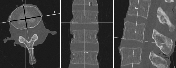
Fig. 2.
Example of a multiplanar view of a human L3 vertebra. From left to right: a transverse, frontal, and sagittal view. In this example, the lower end-plate width and depth were measured in the transverse view. The axes were placed in the correct position in the frontal and sagittal views so that the measurement was performed in the correct anatomical plane

