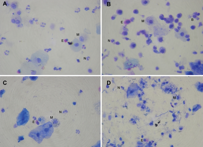
Figure 5).
Comparison of cell morphology (Diff Quik stain, Dade Behring, USA) between fresh sample prepared using the routine method (A), sample preserved in alcohol and prepared by the routine method (B), sample preserved in formaldehyde and prepared using the method of Kelly et al (19) (C), and sample preserved in formaldehyde (D), with moderate debris accumulation. B Bronchial epithelial cell; E Eosinophil; M Macrophage; N Neutrophil

