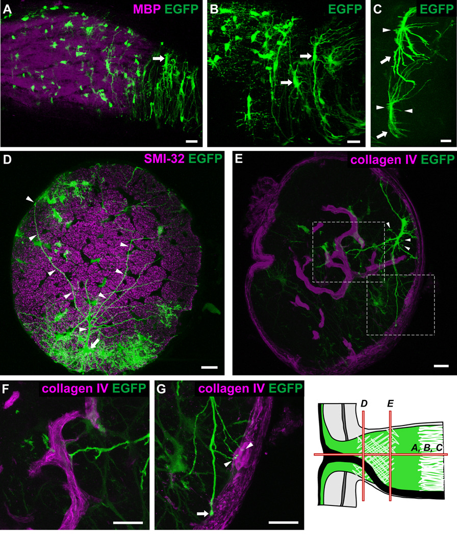
Fig. 5.
Longitudinal and transverse sections of hGFAPpr-EGFP optic nerve showing the morphology of astrocytes within the glial lamina. The schematic of the optic nerve head in the bottom right-hand corner of the figure depicts the origin of the sections shown in each panel. The glial lamina consisted predominantly of transversely oriented astrocytes with thick elongated cell bodies and primary processes extending long distances (A–C; arrows). The overall appearance of these astrocytes was that of a baseball catcher’s mitt. Many small branches emanated from the cell body (C; arrows). Panel D shows an astrocyte (arrow) with processes (arrowheads) that extend the full width of the optic nerve, crossing numerous axon bundles to contact the pial wall via bulbous endfeet. In transverse section, the primary processes extend to contact the pial wall (E, G; arrows) or blood vessels (E, F, G; arrowheads), which have been labeled with collagen IV. Panels F and G represent an enlargement of the area in the dashed square in panel E. Scale bar in each panel = 20 µm.

