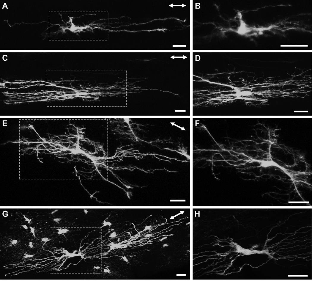
Fig. 6.
Longitudinally oriented astrocytes from the myelinated region of the hGFAPpr-EGFP optic nerve. The astrocytes depicted in this figure were located between the junction of the unmyelinated/myelinated region and approximately three to four millimeters posteriorly. The area represented by the dashed square in panels A, C, E and G are enlarged in panels B, D, F and H. There were numerous morphological varieties of longitudinally oriented astrocytes. Some exhibited very few primary processes that were straight and have few higher order branches (A, B), others possessed undulating processes with many collaterals and small offshoots, giving the astrocyte a ‘hairy’ appearance (C–F). One astrocyte possessed many primary processes, but showed very few higher order branching (G, H). The double-headed arrow in the top right corner of each panel indicates the direction of the long axis of the optic nerve. Scale bar in each panel = 20 µm.

