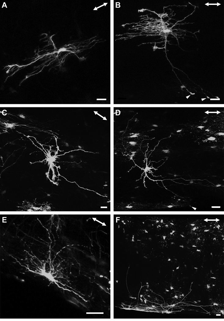
Fig. 8.
There were many morphological varieties of astrocytes, with some projecting primary processes in both the longitudinal and transverse direction (A, B), as well as in random directions (C, D). The primary processes of some of these astrocytes contacted the pial wall with bulbous endfeets (B, D, arrowheads). A gallery of additional examples is shown in the supplementary material (Fig. S2). There were some astrocytes with elongated cell bodies that lay in parallel with the long axis of the optic nerve, adjacent to the pial wall. The astrocytes depicted in this figure were located between the junction of the unmyelinated/myelinated region and approximately three to four millimeters posteriorly. The double-headed arrow in the top right corner of each panel indicates the direction of the long axis of the optic nerve. Scale bar in each panel = 20 µm.

