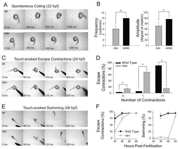
Fig. 1.
nav mutants exhibit abnormal spontaneous coiling amplitude, and diminished touch-evoked behaviors. (A) Top: a 22 hpf a wild type sibling exhibiting a single spontaneous coil. Bottom: an aged matched nav mutant embryo exhibiting a weaker spontaneous coil when compared to wild type sibs. (B) Frequency (left) and amplitude (right) of the spontaneous coils (angle of rotation of the tail) of wild type sib (n=33) were greater than that of nav mutant (n=12) embryos at 21 hpf (t test: p < 0.01 for frequency; p < .05 for amplitude). (C) Top: a 24 hpf wild type sibling touched on the head responds with multiple escape contractions. Bottom: an aged matched nav mutant embryo responds with a single contraction. (D) Percent of touch-evoked escape contractions consisting of no contractions, one contraction and greater than one contraction in wild type (n=30) and nav mutant (n=30) embryos at 24 hpf. All differences (asterisks) were significantly different (t test: p <0.05). (E) Top: a 48 hpf wild type sibling touched on the tail responds with an escape contraction followed by swimming. The embryo appears twice in some frames as the behavior was faster than the video capture rate. Bottom: an aged matched nav mutant responds with an escape contraction but no swimming. (F) Progression of the nav phenotype over the first few days of development compared to wild type. Values represent the average ± SEM escape response displayed by either wild type or mutant embryo groups (n = 3 groups each, 25 embryos each group).

