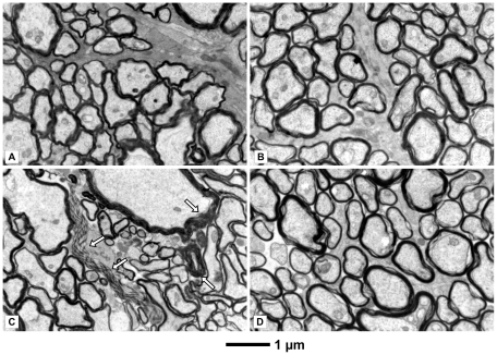
Figure 2. Electron micrographs of the optic nerves displaying the status of myelination.
Rats were injected with rotenone-microspheres or rAAV5-NDI1 or both as described for Figure 1. The optic nerve was examined 1 month after the rotenone infusion. A: control (no injection). B: rAAV5-NDI1-injected. C: rotenone microspheres-injected. Parts of degenerated myelin are indicated by arrows. D: rotenone microspheres-treated with a simultaneous injection of rAAV5-NDI1.

