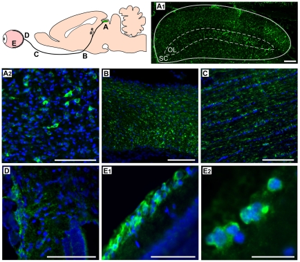
Figure 3. Expression of the Ndi1 protein throughout the optic nerve system.
Rats received rAAV5-NDI1 in the SC. Two months after the injection, tissues were collected from the locations depicted in the cartoon (A – E) and immunohistochemically stained for the Ndi1 protein (green). Nuclei were visualized by DAPI (blue). A1: coronal section of the optical layer (OL) of the superior colliculus (SC). Scale bar = 150 µm. A2: optical layer of the SC. Scale bar = 40 µm. B: sagittal section of the optic chiasma. Scale bar = 100 µm. C: sagittal section of the optic nerve. Scale bar = 100 µm. D: entry point of the optic nerve into the retina. Scale bar = 40 µm. E1: transversal section of the retina. Scale bar = 40 µm. E2: a high magnification image of the retinal ganglion cell layer, Scale bar = 10 µm.

