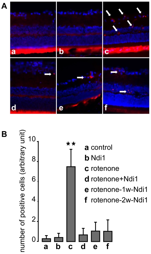
Figure 7. Prevention of apoptosis of cells treated with rotenone by Ndi1 expression.
Rats were subjected to rotenone exposure for 2 months as detailed in the legend to Figure 1 and the retina samples were stained for single strand DNA (red) and DAPI (blue). A: (a) control (no injection), (b) rAAV5-NDI1 injected animal, (c) rotenone microspheres injected animal, (d) rAAV5-NDI1 was injected immediately after the rotenone administration, (e) rAAV5-NDI1 was injected 1 week after the rotenone administration, (f) rAAV5-NDI1 was injected 2 weeks after the rotenone administration. The arrows highlight some of the cells carrying the apoptotic marker. Bright fluorescence seen in the cones and rods layer of the retina is un-specific and is not considered for ssDNA evaluation. B: The number of retinal ganglion cells positive for ssDNA staining was compiled in a histogram. Cells were counted per field of view in different sections separated by at least 100 µm. **p<0.0001, ANOVA test. Error bars represent the mean±SD.

