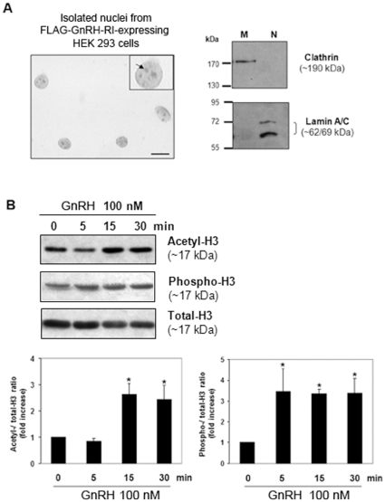
Figure 5. Effect of GnRH stimulation on Histone 3 acetylation/phosphorylation levels in isolated nuclei from HEK 293 transiently transfected with FLAG-GnRH-RI.
(A) Purity assessment of nuclei preparations used in functional assays by bright field microscopy and Western blotting. Left panel: Photomicrograph of a typical nuclei preparation following trypan blue staining. Scale bar = 10 µm. Insert: digital magnification of a single nucleus showing the presence of nucleoles (arrow). Right panel: Immunodetection of the membrane and nuclear markers clathrin and lamin A/C respectively in membrane and isolated nuclei fractions from FLAG-GnRH-R-transfected HEK 293 cells. (B) Time course of Histone H3-Lys9/Lys14 acetylation and -Ser10 phosphorylation in GnRH-stimulated nuclei by Western blot analysis. A representative Western blot is shown. Densitometric values of acetylated and phosphorylated Histone H3 levels were normalized to those of total Histone H3 and the relative intensity ratios were expressed in-fold increase over time zero. Data are means ± S.E.M of four independent experiments. *P<0.05 vs control time zero. Statistical analyses performed using one-way ANOVA followed by Dunnet's ad hoc test.

