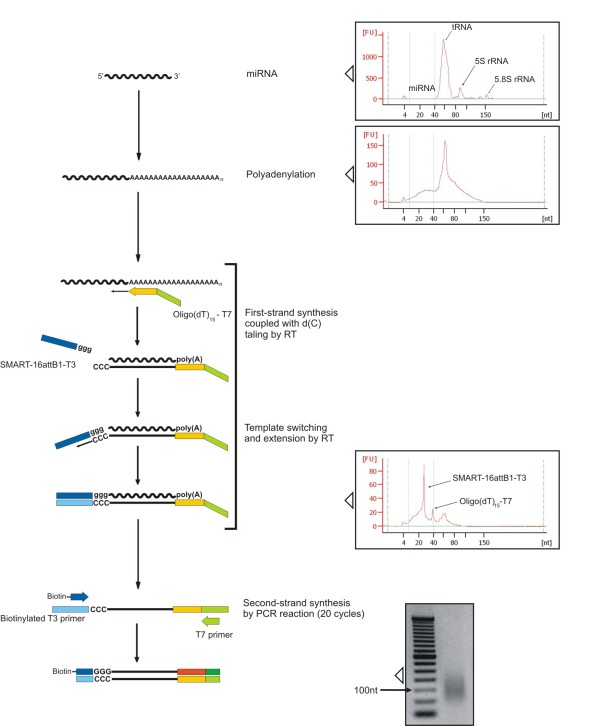
Figure 4.
Schematic representation of miRNA labeling method for bead based quantification. All protocol steps and quality checking with Agilent Bioanalyzer 2100 and gel electrophoresis (2% agarose) are schematically represented: starting material (RNA < 200 nt), the result of polyadenylation reaction with PAP, the synthesis of first-strand cDNA (peaks are associated to Oligo-dT15-T7 and SMART-16attB1-T3 primers) and the second-strand synthesis by PCR amplification.

