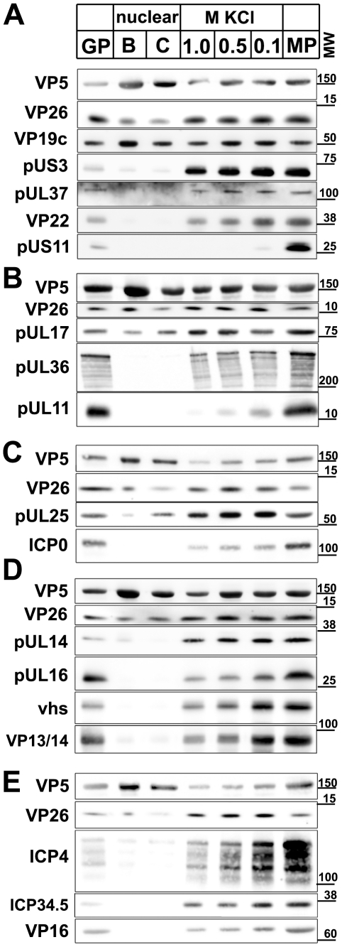
Figure 6. Immunoblot characterization of nuclear and viral HSV1 capsids.
The protein composition of HSV1 capsids (nuclear B capsids, nuclear C capsids, viral capsids treated with 1.0, 0.5 or 0.1 M KCl), virions sedimented from cell culture supernatants (MP, medium pellet) and gradient purified virions (GP) were analyzed by immunoblot using antibodies directed against the capsid proteins VP5 (A–E: pAb NC-1), VP26 (A–E: pAb anti-VP26), VP19c (A: pAb NC-2), pUL17 (B: mAb #203), pUL25 (C: pAb ID1), pUL16 (D: anti-pUL16), or the tegument proteins pUS3 (A: pAb anti-pUS3), pUL37 (A: pAb 780), VP22 (A: pAb AGV30), pUS11 (A: mAb #28), pUL36 (B: pAb #147; anti-middle-pUL36), pUL11 (B: pAb anti-pUL11), ICPO (C: mAb 11060), pUL14 (D: anti-pUL14), vhs (D: pAb 11.388), VP13/14 (D: pAb R220), ICP4 (E: mAb 58S), ICP34.5 (E: pAb anti-ICP34.5) and VP16 (E: pAb SW7). Each blot shows one of three indendent experiments. The panels A, B, C, D, and E represent separate membranes. These data were after a further normalization also integrated into Fig. 3 (white columns).

