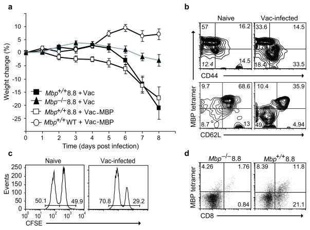
Figure 1.
Wild-type vaccinia virus infection induces autoimmune disease in 8.8 mice. (a) Mbp+/+ 8.8, Mbp−/− 8.8, and wild-type mice (7–11 mice/group) were infected with Vac-MBP or wild-type vaccinia virus on day 0. Mice were weighed daily and the percent weight loss relative to day 0 is shown. The difference in weight loss is significant between MBP+/+ 8.8 and either Mbp−/− 8.8 (P <0.0001) or wild-type mice (P <0.001) infected with wild-type vaccinia virus. Data are compiled from three independent experiments. (b) Splenocytes from 8.8 naïve or wild-type vaccinia virus-infected mice (seven days post-infection) were stained with MBP79-87–H-2Kk tetramer and antibodies specific for CD8, CD44, and CD62L. Flow cytometry analyses are gated on CD8+ cells. Down-regulation of MBP79-87–H-2Kk tetramer staining routinely occurs on activated 8.8 T cells. (c) 8.8 naïve or wild-type vaccinia virus-infected mice (seven days post-infection) were injected with equal numbers of wild-type splenocytes pulsed with MBP79-87 peptide (CFSE-bright) and non-pulsed (CFSE-dim). Mice were sacrificed 20 h later and CFSE-labeled cells analyzed by flow cytometry. Data are representative of two experiments. (d) Mononuclear CNS cells isolated from Mbp+/+ 8.8 and Mbp−/− 8.8 mice seven days post infection with wild-type vaccinia virus were stained with MBP79-87/H-2Kk tetramer and anti-CD8.

