
Fig. 3.
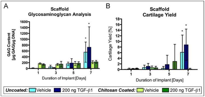
Glycosaminoglycan content and cartilage yield in periosteal cell-laden PCL nanofiber scaffolds. Uncoated and chitosan-coated PCL nanofiber scaffolds were implanted under the periosteum of rabbits for 1, 3, 5, or 7 days followed by six weeks of culture. The implant sites were injected with either 200 ng TGF-β1 or vehicle. (A) GAG content in periosteal cell-laden scaffolds after six weeks of culture (n=7 or 8). (B) Cartilage yield in periosteal cell-laden scaffolds after six weeks of culture (n=16). The data presented are means with 95% confidence interval. The asterisks indicate values that are significantly different than all other time points based on post-hoc testing (p<0.0001).