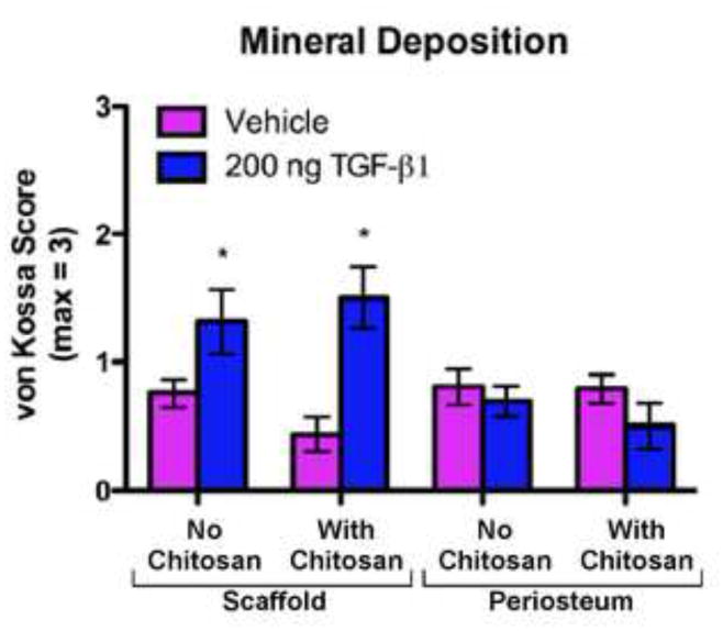
Fig. 6.

Mineral Deposition (based on von Kossa stained histological sections) in uncoated (No Chitosan) and chitosan-coated (With Chitosan) PCL nanofiber scaffolds and periosteum from the implant sites after 7 days of implantation and 6 weeks of culture. The specimens were scored from 0 to 3 (based on a previously validated histological scoring method37) by a blinded technician where 0 = no staining; 1 = partial staining < 50%; 2 = partial staining > 50 %; and 3 = nearly complete or complete staining of the section. The data presented are means with 95% confidence interval (n=16). The asterisks indicate values that are significantly different than the corresponding vehicle control based on post-hoc testing (p < 0.0001).
