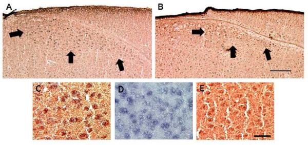
Figure 1.
Double-labeling of SNX (blue) and AR (reddish brown) in the HVC of male (A) and female (B) zebra finches. In A and B, arrows indicate the ventral and lateral borders of the brain region. Panel C depicts double-labeling in the male at higher power. Panels D and E show single-labeld in situ hybridization for SNX2 and immunohistochemistry for AR, respectively, from a different male. Note that labeling, especially for SNX2, tended to be lighter in females. Scale bars = 200 μm for the upper and 25 μm for the bottom panels.

