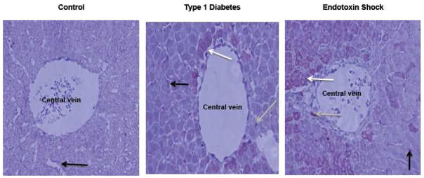
Figure 4. Immunohistochemistry of NOS1 in liver from control, Type-1 diabetic and endotoxin-treated rats.
NOS1 distribution was evaluated in the perivenous area, as shown by immunohistochemistry (at 20X; shown in a red-brown color) (Villanueva et al., 2010, submitted manuscript). Arrows show the distribution of NOS1 in endothelium (black), hepatocytes (white) and Kupffer cells (grey). A rabbit NOS1 polyclonal primary antibody (from Cayman Chemical Co.) was used in the immunostaining procedure that is followed by a diaminobenzidine-based development. In control animals, NOS1 expression was visible only at the endothelium (Control). Using the same conditions in Type-1 diabetic rats, NOS1 was present in hepatocytes, endothelium and Kupffer cells. Nuclear localization of NOS1 was only seen in hepatocytes of Type-1 diabetic rats (compare cells from the three groups). In endotoxin animals (5 h after lipopolysaccharide injection), the number of NOS1-positive cells was higher than that in control animals.

