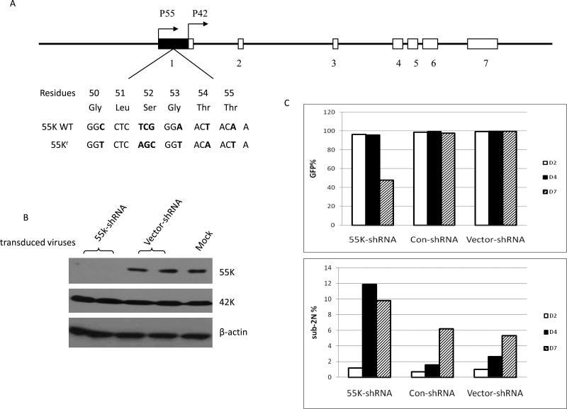
Figure 1. Specific shRNA depletion of 55K CDK9 protein.
A. The genomic structure of the human CDK9 gene and transcription start sites of mRNAs encoding the 55K and 42K proteins are shown (24). The sequence of the wild type 55K mRNA and the sequence of the mRNA encoding the shRNA-resistant 55K protein, termed 55Kr, are shown. B. At four days post-infection, HeLa cell extracts were prepared from cultures infected in duplicate with 55K-shRNA lentivirus, parental Vector-shRNA lentivirus; a single culture was mock-infected. Expression levels of the indicated proteins were examined in immunoblots. C. Expression of the eGFP reporter protein in the cultures infected with the indicated shRNA viruses was examined by flow cytometry at two, four, and seven days post-infection (top panel). Portions of cell cultures were also stained with propidium iodide; the percentage of cells that had a sub-2N DNA content as determined by flow cytometry are shown (bottom panel).

