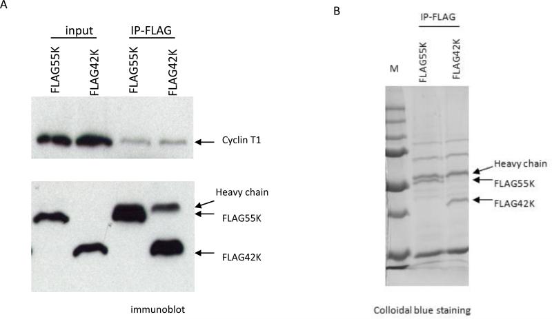
Figure 2. Immunoprecipitations of Flag-55K and Flag-42K CDK9 proteins for proteomic analysis.
A. Plasmid expression vectors for Flag-tagged 55K and 42K CDK9 proteins were transfected into HeLa cells and immunoprecipitations with the Flag antibody were performed at Day 2 post-transfection. An immunoblot was performed to measure the expression levels of the Flag-tagged proteins and the relative amount of Cyclin T1 that co-immunoprecipitated with each protein. B. The plasmid vectors were transfected into HeLa cells and immunoprecipitations with the Flag antibody were performed. A portion of the immunoprecipitate was analyzed on a SDS-polyacrylamide gel which was stained with Colloidal Blue. The remaining portion was subjected to a proteomics analysis to identify proteins present in the precipitations.

