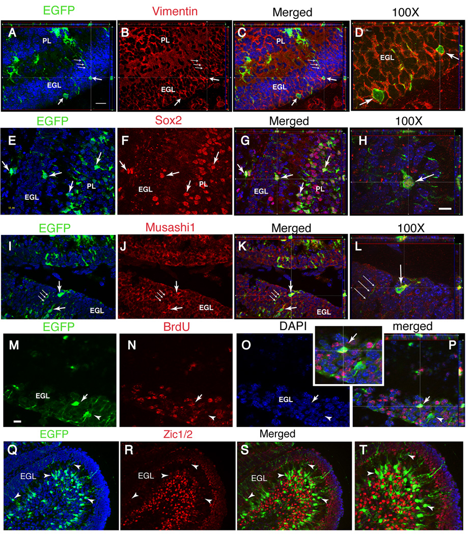
Figure 3. Glial cells with GFAP promoter activity rapidly transition to a GCP phenotype in the EGL.
Double-immunostaining for reporter gene expression and cell specific markers in CGE;CAG-EGFP mice analyzed at P7 after two tamoxifen injections at P5. The antibody markers are indicated above each image. (D,H,L,P,T), higher magnification of (C,G,K,O,S), respectively. The reporter-positive cells in the EGL express vimentin (A–D), Sox2 (E–H) and Musashi1 (I–L). After BrdU incorporation at P5, about 50% of reporter+ cells are BrdU+ (arrows in M–P). In contrast, reporter+ cells do not express the postmitotic GC marker Zic1/2 (Q–T). All panels illustrate apotome 1 µm-thick Z-plane projections. Scale bar in (H) 10 µm for (D,H,L) and 25 µm for (P,T); scale bar in (M) 20 µm for all other panels.

