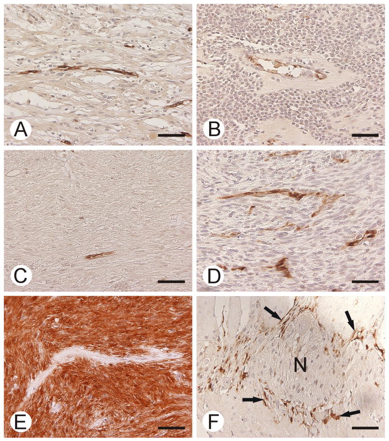
Figure 6.
CA II immunostaining in gastric schwannoma (A), gastric glomus tumor (B), esophageal leiomyoma (C), malignant peripheral nerve sheath tumor (D), GIST (E), and normal smooth muscle (F). GIST shows strong staining for CA II. Capillary endothelium is positive in all other tumor specimens. Note the positive immunoreactions in Cajal cells (arrows in F). Neural myenteric plexus (N) is negative. Bars = 50 μm.

