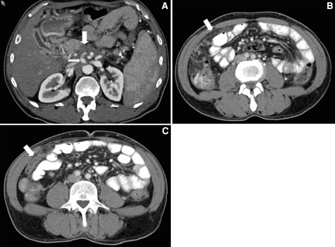
Fig. 6.
Fifty-three-year-old male with unresectable pancreatic adenocarcinoma. A Axial CT image demonstrates a hypovascular mass in the pancreatic body (large arrow) that encases the celiac artery (small arrow). B Axial CT image at the time of diagnosis demonstrates ill-defined soft tissue within the greater omentum (arrow), which was interpreted as either representing peritoneal carcinomatosis or edema. C Axial CT image obtained 3 months later demonstrates nodular soft tissue within the greater omentum (arrow) that is unequivocal for peritoneal carcinomatosis.

