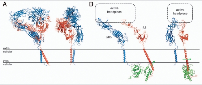
Figure 4.
Integrin bi-directional signaling. (A) Model of a resting integrin receptor. The αVβ3 ectodomain crystal structure was fused to an αVβ3 TM model based on the αIIbβ3 TM complex NMR structure.16,20 The α and β subunits are shown in blue and red, respectively. (B) Model of an active integrin conformation. The α(Calf-1/2) and β(TD/IE2-4) domains of the integrin ectodomain37 are shown dissociated with a schematic active headpiece. The dissociated β TM segment was overlaid with the activating talin2/β1D crystal structure (PDB entry 3g9w). Talin2 is shown in green and the direction of its effected β TM segment reorientation21 is indicated by green arrows. Two orientations, which are related by a rotation of ∼90° about the y axis, are shown for both models.

