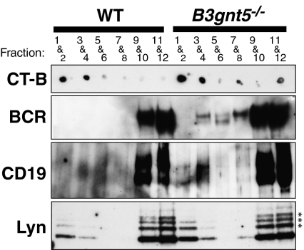
Fig. 4.
Distribution of BCR, CD19, and Lyn in GEMs separated into fractions by sucrose density gradient centrifugation. B cells stimulated with anti-BCR and anti-CD19 Abs were lysed with 1% Triton X-100 solubilization buffer. After sucrose gradient centrifugation, fractions were collected from the top of gradient. Then, every second fraction was combined starting from the top fraction. GM1, a GEM marker, was detected using HRP-conjugated CT-B as shown. After fractionation, immunoprecipitation of BCR and CD19 was performed using anti-BCR and anti-CD19 Abs, respectively. BCR (sIgM) and CD19 proteins in B3gnt5−/− B cells were up-regulated in the GEM fraction (fractions 1–4). Lyn proteins were also up-regulated in the GEM fraction of B3gnt5−/− B cells. The results shown are representative of several independent experiments. *Nonspecific bands.

