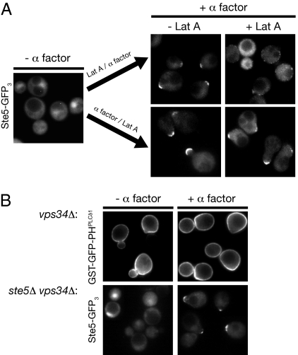
Fig. 4.
Membrane recruitment and shmoo tip localization of Ste5 do not require actin filament function or PtdIns 3-kinase Vps34 activity. (A) Exponentially growing cultures of ste5Δ cells harboring a low-copy (CEN) vector expressing from the STE5 promoter Ste5-(GFP)3 (Left) were either not treated (−) or treated (+) with 100 μM latrunculin A (Lat A) for 15 min and then treated with 20 μM α-factor for 30 min (Right, Upper) or treated with 20 μM α-factor for 30 min and then either not treated (−) or treated (+) with 100 μM Lat A for 30 min (Right, Lower). Cells were then examined by standard epifluorescence microscopy; each panel depicts a collage of representative cells. (B) Upper, PtdIns 3-kinase Vps34 is not required to generate PtdIns(4,5)P2 anisotropy. Expression of the GST-GFP-PHPLCδ1 probe was induced and shut off in a vps34Δ mutant as described in Fig. 1A, and the cells then were either not treated (−) or treated (+) with 5 μM α-factor for 60 min and examined by standard epifluorescence microscopy. Lower, PtdIns 3-kinase Vps34 is not required for plasma membrane recruitment of Ste5. Exponentially growing cultures of a ste5Δ vps34Δ double mutant harboring a low-copy (CEN) vector expressing from the STE5 promoter Ste5-(GFP)3 were either not treated (−) or treated (+) with 20 μM α-factor for 90 min and examined by epifluorescence microscopy. Each panel depicts a collage of representative cells.

