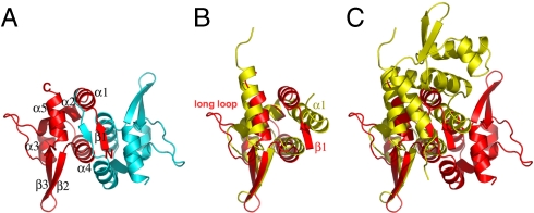
Fig. 1.
B. thuringiensis pBtoxis TubR structure. (A) One TubR subunit is red and the other cyan. Secondary structural elements and N and C termini are labeled. (B) Superimposition of one subunit of TubR (Red) onto a S. aureus CzrA subunit (Yellow). Regions with different structures are labeled. (C) Same superimposition as B showing the location of the other subunit in the TubR and CzrA dimers after one subunit is overlaid. A–C are in the same orientation to highlight differences. Figs. 1 A–C, 2 C and D, 3 A, C, and E, 4B, and 5B were made by using PyMOL (31).

