Fig. 4.
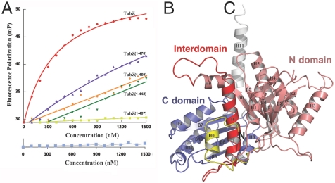
TubZ interacts with TubR-DNA and contains a tubulin/FtsZ fold. (A) FP assay measuring binding of FL TubZ, TubZ(1-470), TubZ(1-460), TubZ(1-442), and TubZ(1-407) to TubR-DNA. Below is the control (TubZ titrated into DNA alone). Millipolarization units and TubZ concentration (nM) are along the y and x axes, respectively. (B) TubZ(1-428) structure. The N domain or GTP-binding domain is colored salmon and the C domain purple. The interdomain helix, H7, is red. TubZ also contains an N-terminal helix, H0 (Yellow), and a C-terminal helix, H11 (White).

