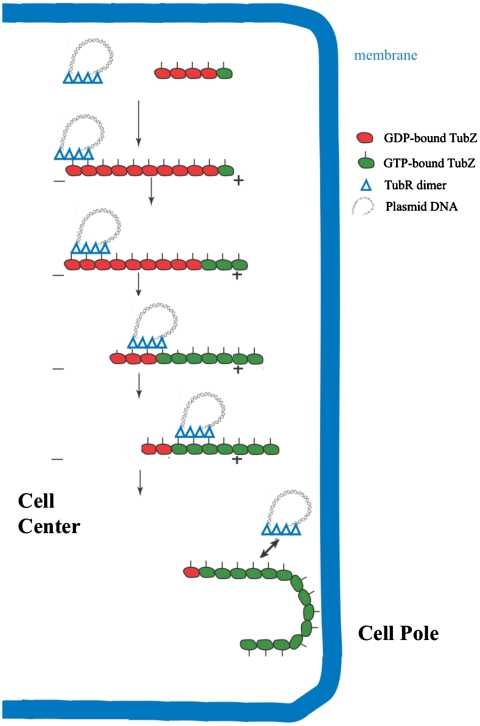
Fig. 6.
pBtoxis DNA partition model. In the first step, TubR, which is bound to its centromere on one of the replicated pBtoxis plasmids, contacts the TubZ C-terminal region (indicated by lines pointing from the TubZ “circles”) in a treadmilling TubZ polymer. TubZ subunits are lost from the - end and are added to the + end. TubR is pulled along the growing polymer by its TubR-TubZ interaction until it reaches the cell pole and is knocked off when it comes into contact with the membrane at the cell pole. TubZ reverses direction and may pick up the other TubR-pBtoxis complex and deliver it similarly to the opposite cell pole.

