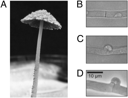
Fig. 3.
Photograph and micrographs of C. cinerea. (A) Mature C. cinerea fruiting body that is shedding spores. The upper surface of the cap has loosely adhering “veil cells.” The lower surface of the cap is composed of “gills” which support the basidia (meiotic cells). The cap is elevated several centimeters above the Petri dish by the “stipe” (stalk). (B) Simple septum between two cells in a monokaryotic hypha. (C) “False clamp” between two cells in an “A-on” hypha. (D) True clamp connection between two cells in a dikaryotic hypha (“A-on B-on”). Magnification in B–D is the same.

