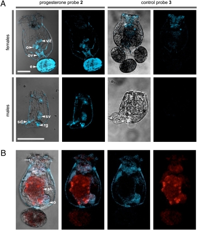
Fig. 2.
Imaging studies with the rotifer Brachionus manjavacas. (A) Confocal fluorescence microscopic images depicting rotifers treated with progesterone probe 2 or control probe 3. (B) Two-color confocal images showing a female rotifer fed T. suecica then treated with 2; T. suecica autofluoresces in the red channel, and 2 fluoresces in the blue channel. (Scale bars: 100 μm.) Blue and red fluorescence were collected with excitation at 375 ± 10 nm and emission at 458 ± 10 nm and excitation at 488 ± 10 nm and emission at 518 ± 10 nm, respectively. Organelles are noted by: vit, vitellarium (yolk gland); o, ovaries; ov, oviduct; e, egg; sv, seminal vesicle; rg, rudimentary gut; st, stomach; sd, sperm duct.

