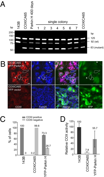
Fig. 5.
Partially reverted COXICA65 expressing YFP-Parkin (Parkin H) contains a mixed population. (A) 143B, COXICA65, Parkin H cybrid cells following transfection for 400 d (200 d in the absent of FACS selection) and seven single colonies isolated from the Parkin H cells in Fig. 4E were analyzed for the ratio of wild-type and mutant mtDNA by PCR-RFLP. (B) The COXICA65 cells Parkin-enriched for wild-type mtDNA [Parkin H, 67 d postenrichment (Fig. 4E)] were fixed and stained with Tom20 antibody (mitochondria, blue) and COXI antibody (red). YFP-Parkin is green. Cells with neither YFP-Parkin nor COXI signal were circled. Cells without YFP-Parkin but with COXI signal were marked with *. (Scale bar, 20 μm.) (C) The percentage of COXI-positive and -negative cells were scored in untransfected 143B, COXICA65 cybrid cell lines and COXICA65 cybrid cells enriched to 90% wild-type mtDNA by YFP-Parkin expression, followed by 67 d release from Parkin selection (Parkin H 67 d postenrichment) (A, Upper) considering only YFP-Parkin-negative cells. More than 110 cells lacking detectable YFP-Parkin signal were counted in each sample. (D) Cytochrome c oxidase activity (COX) assay. COX activity for each sample is reported relative to 143B, which contains 100% wild-type DNA.

