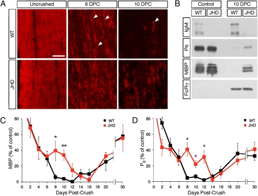
Fig. 1.
Myelin clearance is delayed in B-cell–deficient mice. (A) Staining of paraformaldehyde-fixed cryosections obtained from control and crushed sciatic nerves at 6 and 10 d postcrush (DPC) using the FluoroMyelin dye (red) at a dilution of 1:300. Arrowheads mark degenerating myelin ovoids. (B) Western blot of lysates obtained from WT and JHD sciatic nerves probed with anti-IgM, anti-P0, anti-MBP, and anti-FcR common γ chain antibodies. (C) Quantification by Western blotting of MBP levels in WT and JHD sciatic nerve lysates both following sciatic nerve injury and in uncrushed control nerves. (D) Quantification of P0 levels in WT and JHD mice after injury. Total protein was assessed by the BCA assay and each well was loaded with the same amount of total nerve protein. All data are from male mice and are presented as mean ± SEM, n = 4–10 animals per genotype per time point (*P < 0.05; **P < 0.01). (Scale bar, 200 μm.)

