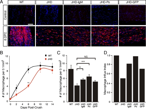
Fig. 3.
B-cell deficient mice have delayed macrophage influx following sciatic nerve injury that is rescued by passive antibody transfer. (A) Cryosections of uninjured and injured sciatic nerves from WT and JHD mice following passive Ig transfer immunostained with the combined macrophage-specific markers CD68 and F4/80 antibodies (shown in red). DAPI was used as nuclear counterstain (blue). (B) Time course of macrophage influx into the nerve after crush. (C) Quantification of macrophage numbers in JHD mice at 6 d postcrush (DPC) following 1 dose of passive transfer of Ig at 2 DPC. (D) Macrophage influx index in JHD nerves. Rate of macrophage influx in JHD nerves between postcrush day 2 and day 6 normalized to WT nerves (as measured from slope of the line between days 2 and 6 in B) in mice following 1 dose of passive transfer of Ig at 2 DPC. All data are from male mice and are presented as mean ± SEM, n = 7 animals per genotype per time point (*P < 0.05; **P < 0.01). (Scale bar, 200 μm.)

