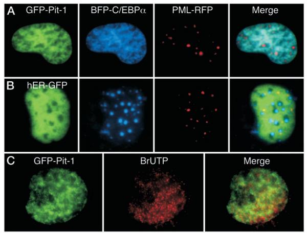
Fig. 4.
Pit-1 Recruits the Coexpressed C/EBPα
GHFT1–5 cells were cotransfected with BFP-C/EBPα, PML-RFP, and either GFP-Pit1 (panel A) or GFP-ER (panel B). Sequential images from the same focal plane were acquired using suitable filters as described in Materials and Methods. The images were merged to show regions of overlap, with blue and green overlap indicated by cyan color, and regions of red and green overlap indicated by yellow color in the merged image. C, GHFT1–5 cells expressing GFP-Pit1 were permeabilized and exposed to BrUTP for 20 min. BrUTP was then immunohistochemically detected in fixed cells with a Texas red-conjugated secondary antibody as described in Materials and Methods. Dual color images were obtained in the same focal plane, and the images were merged to show regions of overlap, which appear yellow in the merged image.

