Fig. 5.
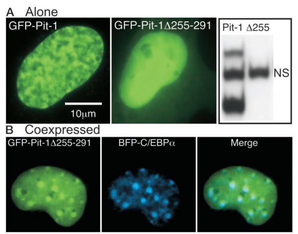
The HD of Pit-1 Is Necessary for Specific Subnuclear Interactions with C/EBPα
A, Fluorescence images of living GHFT1–5 cells expressing either GFP-Pit-1 (left panel) or GFP-Pit1 lacking the carboxyterminal portion of the HD (GFP-Pit-1Δ255–291, middle panel). Cell extracts were prepared from HeLa cells expressing either GFP-Pit-1 or GFP-Pit-1Δ255–291 and samples were incubated with a labeled PRL 1P Pit-1 response element as described in Materials and Methods. EMSA showed that GFP-Pit-1 (lane 1) bound to the Pit-1 DNA element resolved as two distinct complexes, whereas there was no detectable binding of GFP-Pit1Δ255 (lane 2) to the 1P Pit-1 DNA element (NS, nonspecific, see Fig. 2). B, Dual color fluorescence images of living GHFT1–5 cells coexpressing GFP-Pit1Δ255 and BFP-CEBPα were acquired from the same focal plane. The images were merged to show regions of overlap, which appear as cyan color in the merged image.

