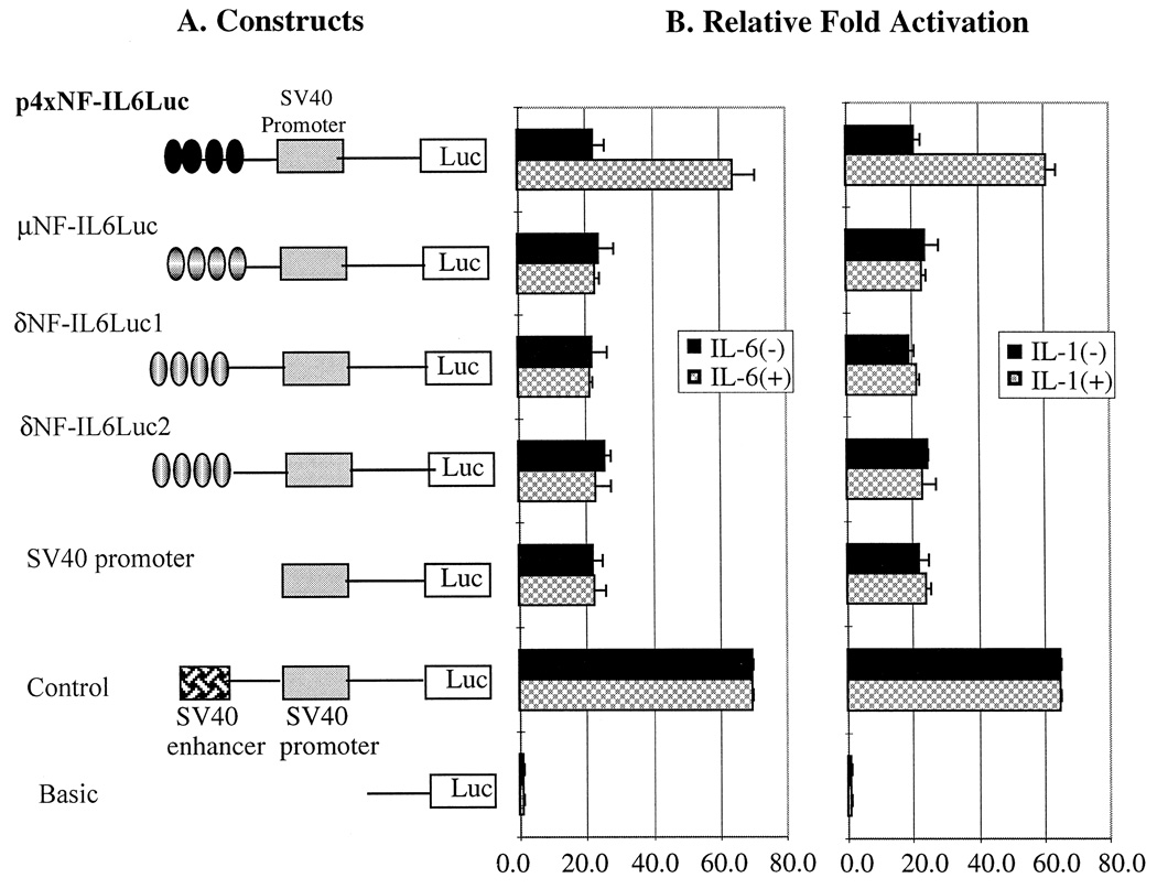
Fig 3.
Construction of four tandemly repeated core sequences of the cytokine response elements (NF-IL6 binding sites) from opioid receptor promoter region (A) and the fold activation measured by luciferase activities after transfected into U266 and Raw264.7 with (+) and without (−) cytokine treatment (B). The cytokines used for this experiment are IL-1 α + β (200 U/ml) and IL-6 (500 U/ml) and incubated for 72 h. A reference plasmid, p4 × NF-IL6Luc, which contains four copies of NF-IL6 binding sites of IL-6 gene, was used as a positive control for cytokine responses. There was no significant difference in luciferase activity between two different cell lines, U266 and Raw264.7 in terms of the represents both cell line, U266 and Raw264.7. Time course and dose-dependency experiments showed no different promoter activity from shown in this figure. (· · · ·) Indicates four tandem repeats of NF-IL6 binding sites. Three times of individual experiments were performed with duplicates each time.

