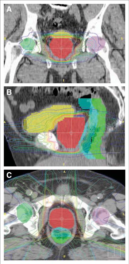
FIGURE 1.
Sample Treatment Plan for External Beam Radiation Therapy to the Prostate. (A) Coronal, (B) sagittal, and (C) axial computed tomographic images of a representative plan for low-risk prostate cancer. Isodose lines depict radiation doses at various distances from the target tissue. Full dose is contained within the pink isodose line. Shading of organs of interest is as follows: prostate (red), bladder (yellow), anterior rectum (light blue), and posterior rectum (green). The close proximity of bladder and anterior rectum to the prostate leads to significant radiation doses to portions of these structures.

