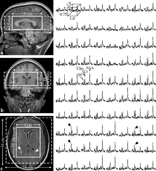
Fig. 1.
Left: Sagittal (a) and coronal (b) T1-weighted and axial T2-weighted FLAIR (c) MRI of patient #12 in table 1, with the VOI and FOV (solid and dashed white frames) superimposed. Note characteristic periventricular hyperintensities on c (arrows) and little or no atrophy.
Right: Real part of the 8×10 (LR×AP) 1H spectra matrix from the VOI on c, on common frequency (1.4 to 3.8 ppm) and intensity scales. Note the spectral resolution and SNR in these 0.75 cm3 voxels and elevated Cho and mI at the lesions (black arrows).

