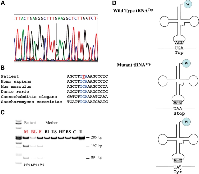
Figure 2.
(A) Sequence of the mitochondrial tRNATrp gene in DNA isolated from the patient's muscle. The arrow indicates the C5545T mutation. (B) Alignment of sequences of the anticodon loop of tRNATrp from different eukaryotic species. (C) PCR–RFLP analysis of the mutation in different tissues of the patient and of his mother. Radiolabeled fragments, after digestion with DraI, were separated on a gel, visualized and quantitated using a BioRad phosphor imager. The wild-type yields a single fragment of 286 bp, the mutant two fragments of 197 and 89 bp. In red patient tissues. M, skeletal muscle; BL, blood leukocytes; F, hair follicles; US, urinary sediment; HF, hair follicles; BS, buccal smear; C, healthy control; U, uncut fragment. (D) Schematic representation of the structure of wild-type and mutant tRNATrp and the possible consequences of the mutation in the anticodon on codon recognition.

