
Fig. 8.
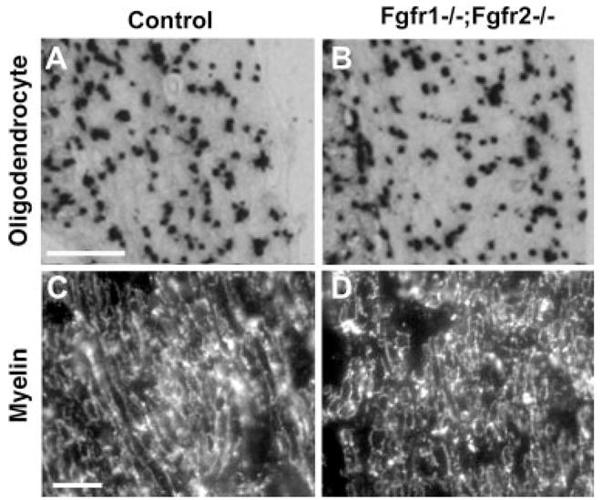
Oligodendrocytes and central myelin appear to be similar in Fgfr1−/−; Fgfr2−/− and control mice. Transverse sections of the ventral cochlear nucleus (A,B) or central segment of cochlear nerve (C,D) from control and mutant mice were analyzed by in situ hybridization for the oligodendrocyte marker PLP mRNA (A,B) or by immunohistochemistry for myelin marker MBP (C,D). Expression patterns of PLP mRNA and MBP were similar in control and Fgfr1−/−; Fgfr2−/− mice. Analysis of three or four mice of each genotype, ranging between 4 and 8 months of age, gave similar results. Representative images are shown. Site at which sections were cut as referenced in Figure 2, A,B: 5; C,D: 4. Scale bars = 200 μm in a (applies to A,B); 10 μm in c (applies to C,D).