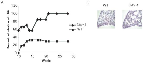
Figure 6. Caveolin-1 deficient mice are readily colonized with P. aeruginosa strain N6.
A. cav1 KO (Cav-1) and WT mice (N=10) were exposed to P. aeruginosa strain N6 in the sterile drinking water for one week, subsequently placed on acid water and monitored for oropharyngeal colonization. Seven weeks later the mice were treated with the antibiotic meropenem in the drinking water for two weeks and subsequently reinstated on acid water. Animals were monitored for the presence of P. aeruginosa in the throat by swab cultures after the antibiotic treatment was withdrawn. The percent of oropharyngeal colonization of cav1 KO and WT mice at each week is plotted. Differences in the probability of colonization of cav1 KO mice versus WT mice are significant at a level of P<0.001 using generalized estimating equations (26). B. Lung sections harvested from chronically colonized cav1 KO or WT mice and stained with H&E. Images shown are representative examples of infected lungs from Cav-1 deficient and WT animals.

