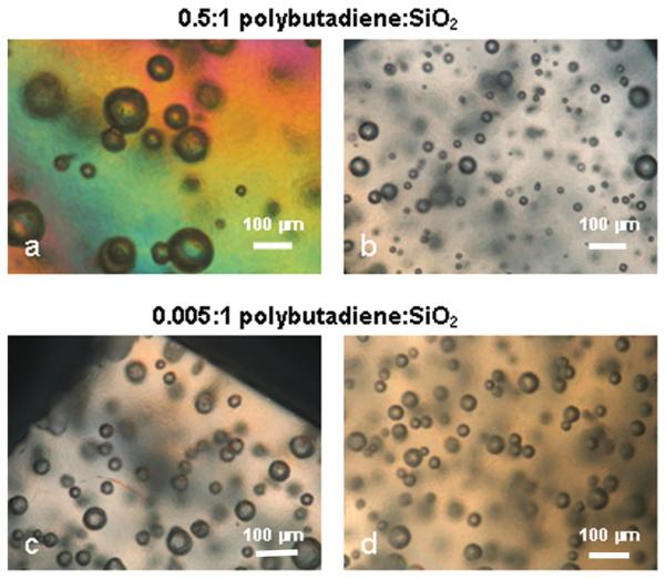
Figure 7.

Optical microscope images near (a,c) and far (b,d) from fracture surfaces. Note extensive shear damage and markedly increased size of aggregates, possibly due to cavitation, in (a). Much less shear damage is evident in (c). Some planes are out of focus. Examined under crossed polarizers. Crosshead speed – 0.001mm/min.
