Figure 1.
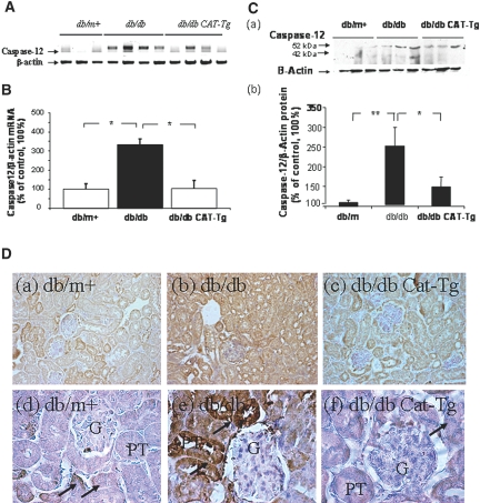
Caspase-12 expression is validated in mouse kidneys at week 20. (A) Southern blotting of conventional RT-PCR analysis of caspase-12 mRNA expression in RPTs of db/m+, db/db, and db/db CAT-Tg mice. (B) RT-qPCR analysis of mouse caspase-12 mRNA expression in RPTs of db/m+ (wt), db/db, and db/db CAT-Tg mice. Caspase-12 mRNA levels in db/m+ mice were considered as 100%. Each point represents the mean ± SD of six animals. *P < 0.05; **P < 0.01. (C, a) Immunoblotting (antibody from Cell Signaling) of mouse caspase-12 protein expression in RPTs of db/m+ (wt), db/db (db), and db/db CAT-Tg (db-Cat) mice. (b) Densitometry of the data in a. (D) Immunohistochemical staining for caspase-12 in db/m+, db/db, and db/db CAT-Tg mouse kidneys, using rabbit anti–caspase-12 polyclonal antibodies (antibody from eBioscience). Arrows indicate caspase-12 immunopositive cells in proximal tubules. Magnifications: ×200 in D, a through c; ×600 in D, d through f.

