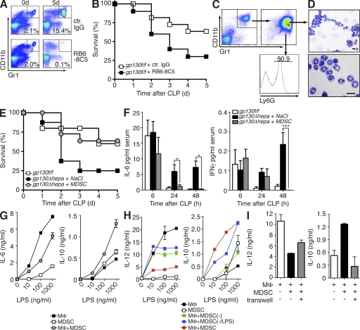
Figure 3.
MDSCs regulate inflammatory responses and confer protection during sepsis. (A and B) gp130f/f mice were injected with monoclonal antibodies against Gr1 (RB6-8C5) 24 h before and after CLP. Efficient depletion of Gr1+ MDSCs was determined by flow cytometry of total splenocytes (A). Survival was monitored (B; n = 9 per group; pooled data of three independent experiments). (C) gp130f/f donor mice underwent CLP and 5 d later MDSCs were isolated from the spleens. CD11b+Gr1+ purity of >90% was confirmed by flow cytometry (top right). Further analysis of Ly6G expression of MDSCs (bottom right). (D) Cytospins of MDSCs. Bars, 10 µm. (E) gp130Δhepa mice received either 5 × 10⁶ MDSCs or saline 1 and 24 h after CLP and survival was determined (n = 12 per group; pooled data of three independent experiments). (F) Serum samples were taken at the indicated time points and analyzed for IL-6 and IFN-γ by ELISA. (G) Peritoneal macrophages (MΦ) were co-cultured with MDSCs isolated from septic gp130f/f mice at a ratio of 1:3 and were stimulated with LPS at the indicated concentrations. Cytokine release was determined by ELISA (one out of five independent experiments shown). (H) Gr1+CD11b+ cells were isolated from the spleens of septic mice (MDSC) or naive mice (MDSC(−)) and co-cultured with macrophages and stimulated with LPS. Some cells from naive mice were pretreated with 100 ng/ml LPS in vitro before co-culture with macrophages (MDSC(−/LPS)). (I) MDSCs and macrophages were separated in a Transwell and stimulated with LPS. Cytokine release was determined by ELISA (H and I; one out of three independent experiments shown). *, P < 0.05; ***, P < 0.001. Data are presented as means ± SEM.

