Figure 5.
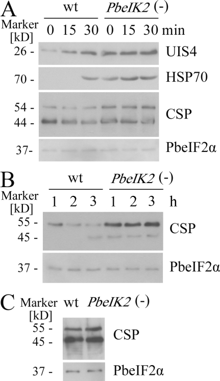
Enhanced liver stage proteins expression in PbeIK2 (-) salivary gland sporozoites. (A) Wild-type and PbeIK2 (-) sporozoites were incubated at 37°C for different times, then UIS4, HSP70, and CSP were visualized on immunoblots by using the polyclonal UIS4 antisera, mAb 2E6, and 3D11, respectively. Antiserum against total PbeIF2α served as loading controls. (B) Sporozoites were incubated at 25°C for different times, and the amounts of CSP secreted into the culture medium were immunoblotted with mAb 3D11. (C) CSP in wild-type and PbeIK2 (-) midgut sporozoites. On day 14 after the first infectious blood meal, wild-type and PbeIK2 (-) parasites were dissected from mosquito midguts, and CSP was visualized on immunoblot. Immunoblots were repeated at least three independent experiments.

