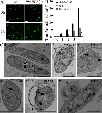
Figure 7.
Morphology of PbeIK2 (-) salivary gland sporozoites. (A) Fluorescence view of wild-type and PbeIK2 (-) sporozoites fixed immediately after dissection from mosquito salivary glands or after 4 h incubation at 37°C. Bars, 10 µm. (B) Sal treated wild-type sporozoites were incubated at 37°C for different hours. Numbers of transformation bulbs were counted. One group of sporozoites was incubated with 50 µM proteasome inhibitor MG132 for 4 h. These data were obtained from two independent experiments. (C) TEM of PbeIK2 (-) sporozoites fixed immediately after dissection from mosquito salivary glands. Mn, Micronemes. Panels i and ii, wild-type; iii-vi, mutant. Arrows in panels iii and iv point to bulbs; in panel v, membrane detachment; in panel vi, transformation bulb with redistribution of organelles. Bars, 1 µm.

