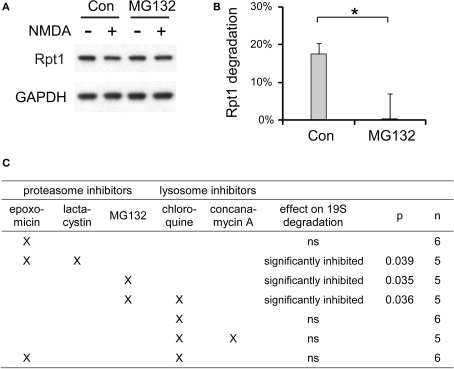
Figure 10.
The degradation of 19S particles is proteaosme-dependent. (A) Proteasome inhibitor MG132 (40 μM) was added to cultured hippocampal neurons 30 min before NMDA stimulation (20 μM, 3 min). After NMDA washed-out, MG132 was applied again for 4 h before neuronal lysates were collected. In control (Con) experiments, neurons were only treated with NMDA. Rpt1 levels were determined by SDS-PAGE and immunoblotting. GAPDH served as loading control. (B) 19S degradation induced by NMDA in (A) was quantified by densitometry (n = 5, mean ± SEM). Statistical significance (*p < 0.05) was determined by t-test assuming unequal variance. (C) The same experiments as in (A) and (B), performed with different combinations of proteasome inhibitors (2 μM epoxomicin; 10 μM lactacystin; 40 μM MG132) and lysosome inhibitors (150 μM chloroquine; 100 nM concanamycin A). Statistical significance was determined using t-test (p-value is listed, or ns for not significant, p > 0.05).

