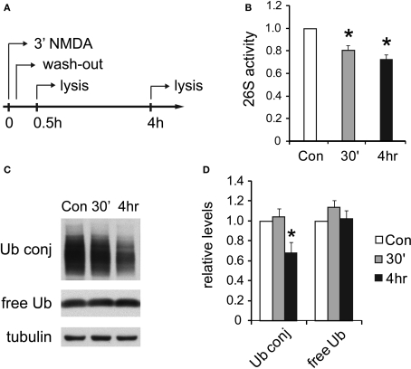
Figure 6.
UPS activity is reduced after NMDA treatment. (A) Cultured hippocampal neurons were treated with 20 μM NMDA for 3 min, a standard procedure for inducing chemical LTD. Whole-cell lysates were prepared at 30 min or 4 h post-treatment. (B) Decreases in proteasome activity after NMDA exposure. 26S proteasome activities in neuronal lysates were assayed with Suc-LLVY-AMC (see Materials and Methods). NMDA-treated samples (30’ and 4 h) are normalized to the untreated control (Con) (mean ± SEM, n = 5, *p < 0.05 by paired t-test). (C) Ubiquitin conjugates decrease after NMDA exposure. Neuronal lysates resolved by SDS-PAGE were blotted using antibodies against ubiquitin-protein conjugates (Ub conj) and free monomeric ubiquitin (free Ub). Tubulin served as the loading control. (D) Signals in (C) quantified by densitometry (n = 5, *p < 0.05 by paired t-test).

