Fig. 6.
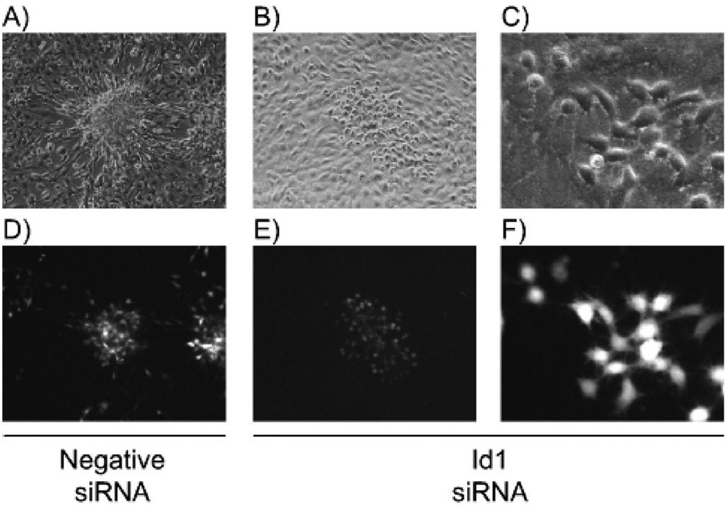
Morphology of LMP1 transformed cells. Rat-1 cells were transfected with negative control (panels A and D) or Id1 siRNA (panels B, C, E, and F) in twelve-well plates. The following day cells were transduced with LMP1-expressing retrovirus and focus formation assays were performed for 7 days. Monolayers were examined by phase contrast (panels A–C) and fluorescence microscopy (panels D–F) for focus formation at 10× low power (A, B, D, and E) and 40× high power (C and F). Representative micrographs are depicted.

