Figure 1.
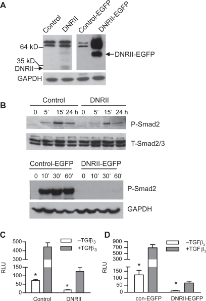
Effects of DNRII expression on TGFβ signaling in HEC-1-A cells. A) Expression of DNRII or DNRII-EGFP were detected with Western immunoblotting. GAPDH levels were used to indicate equal sample loading. B) Exponentially growing cultures of the indicated cells were treated with 1.0 ng/ml of TGFβ3 for different time periods as indicated. Cell lysates were collected for measuring the levels of phosphorylated Smad2 (P-Smad2) and total Smad2/3 with Western immunoblotting as described in the Materials and methods. (C and D) The indicated cells were plated in 12-well plates and transiently transfected with pSBE4-Luc and β-gal constructs with Fugene6. The transfected cells were treated with or without 1.0 ng/ml TGFβ3. The activities of luciferase and β-gal were measured 24 h later. Luciferase activity was normalized to β-gal activity and expressed as relative luciferase unit (RLU). The results plotted represent the means ± SEM from triplicate transfections.
Notes: *Denotes significant (P < 0.05) difference between control and its respective DNRII cells in the absence of exogenous TGFβ3.

